Filarial Nematodes
Filarial Nematodes
Editor: Edward Mitre
Filariae are tissue-invasive nematodes that cause diseases such as elephantiasis, river blindness, and eye worm. The term “filaria” derives from the latin word “filum” that means “thread”, alluding to the thread-like appearance of these worms. Unlike intestinal nematodes, the adult forms live and reproduce in specific host tissues (NOT in the intestine) and produce larval forms called microfilariae. Filarial nematodes are transmitted through the bites of arthropods (mainly mosquitoes and flies).
Although we should not have bias, building the filariasis module was one of our favorites because these worms have very complex biology and unique features. We hope you enjoy learning it as much as we enjoyed making this lesson!
Section Contents
Lymphatic filariasis
Background & Epidemiology
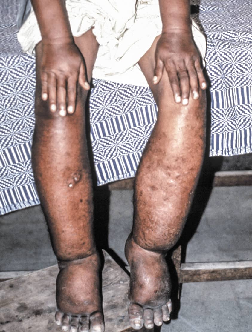
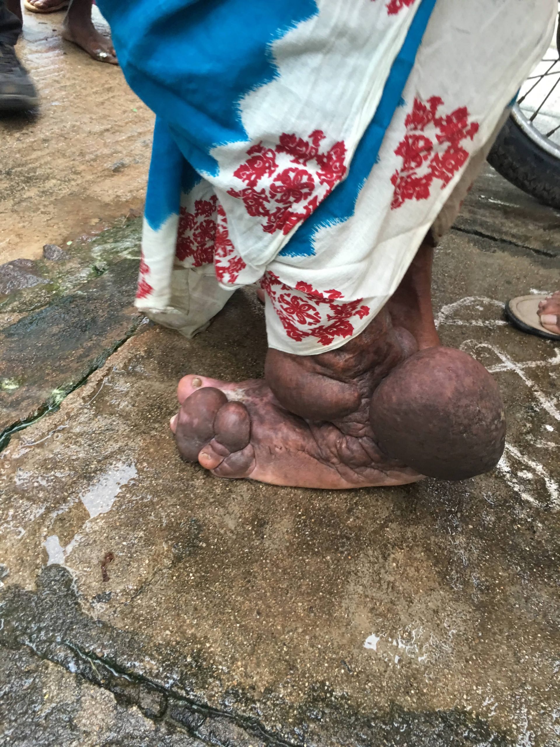
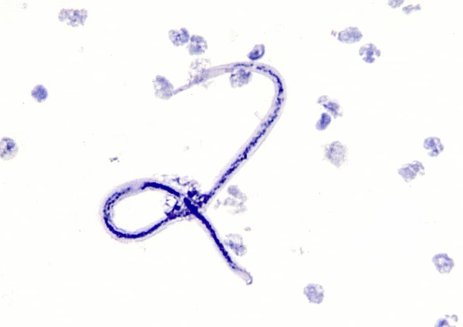
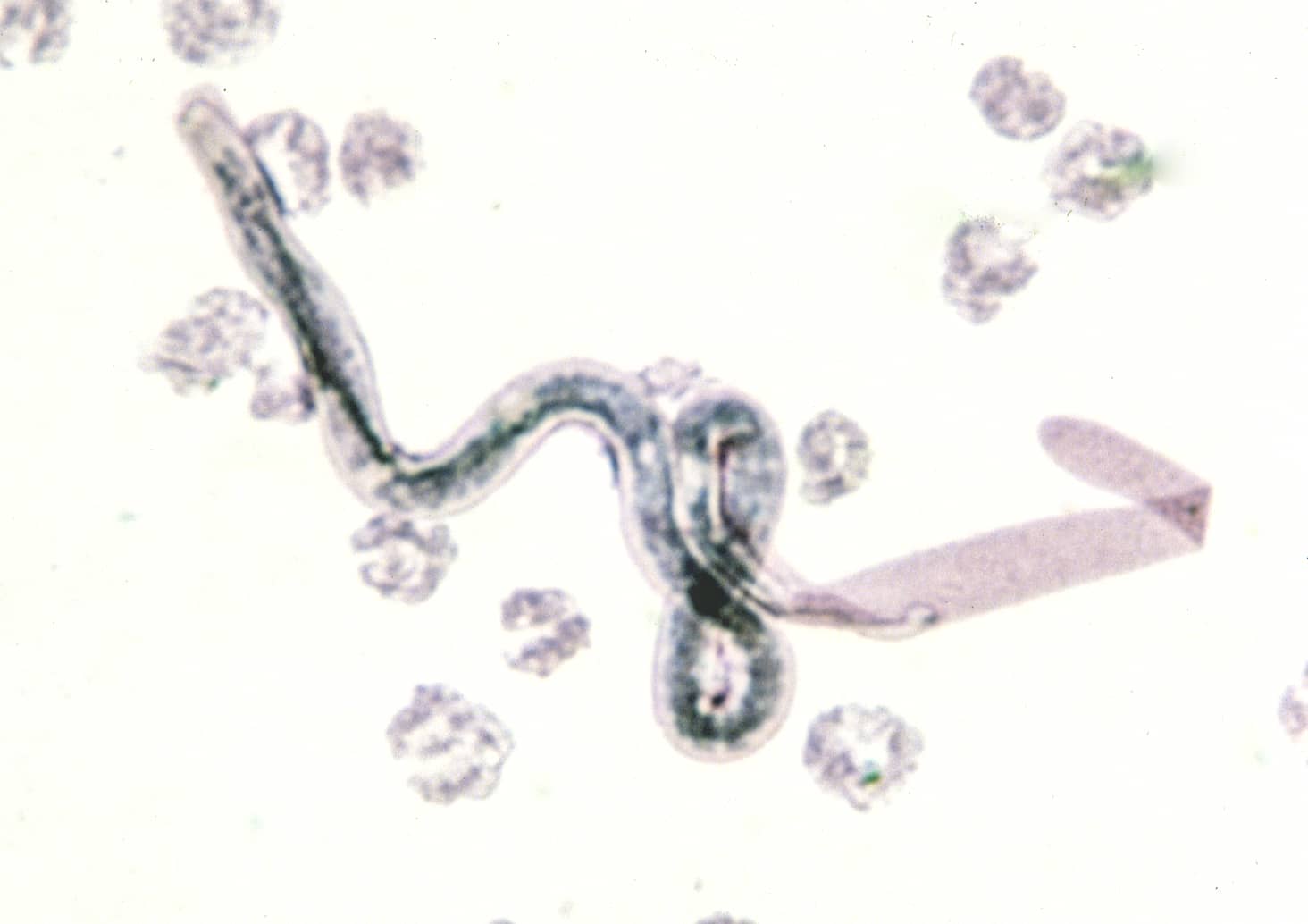
The first group of filarial worms we will describe are Wuchereria bancrofti and Brugia malayi. Third stage larvae of these worms are transmitted to people by mosquito bites, and adult worms live in the lumen of the lymphatic vessels. The disease caused by these worms is called lymphatic filariasis (LF), which is characterized by lower extremity swelling. W. bancrofti is responsible for close to 90% of LF infections, whereas B. malayi causes about 10% [rarely, LF can be caused by other species such as Brugia timori, which will not be described in this lesson]. Infection with either requires repeated exposure (typically >3 months) to infected mosquitoes (rare in travelers).
Although both Wuchereria and Brugia share similar life cycles and pathogenesis, they have key differences that you should remember - summarized in the table below.
| W. bancrofti | B. malayi | |
| Mammalian reservoir | Humans (Anthroponosis) | Felines and Monkeys (Zoonosis) |
| Vectors | Culex spp., Anopheles spp, Aedes spp. | Mansonia spp., Anopheles spp. |
| Distribution | Tropical regions of SubSaharan Africa, Egypt, Southeast Asia, Indian subcontinent, Brazil, Guyana, Dominican Republic, Haiti and Southwest Pacific Islands | Malaysia, Indonesia, Philippines |
")
Figure 1. Age-standardized DALY rates for Lymphatic filariasis (taken from Global Health Metrics, Open Access)
Life Cycle
The life cycle of both Wuchereria and Brugia is very similar, explained in this video:

Figure 2. Life cycle of Wuchereria bancrofti
Pathogenesis & Clinical Presentation

Diagnosis
As with all diseases, diagnosis starts with clinical suspicion in the right epidemiological context (ask yourself what is the presentation? And who is the host?). Definitive diagnosis can be supported through different modalities based on the disease stage:
Treatment
Treatment of filarial nematodes is COMPLICATED. Many physicians are unfamiliar with these drugs, and they need careful management and monitoring. You will NOT treat this alone.
As a general concept please know the following:
- The drug of choice for lymphatic filariasis (W. bancrofti, B. malayi) is called diethylcarbamazine (DEC), which kills microfilariae AND adult worms.
- Before starting DEC, one MUST rule out co-infection with Onchocerca volvulus and Loa Loa! Why? Because DEC can rapidly kill these worms and causes blindness, encephalopathy and other side effects you don’t want to run into.
- There are other alternative regimens that include the use of albendazole, ivermectin or doxycycline - phone a friend first!
- In addition to antiparasitics, individuals will need care for some of the complications. For example, they may need surgery for the hydrocele, wound care for lymphedema, compression bandages and garments, among others.
Prevention
- Mass drug administration (MDA) campaigns to interrupt transmission in endemic areas. Regimens vary per region, but include single doses or combinations of albendazole, ivermectin, and DEC.
- Mosquito control and mosquito prevention
Onchocerciasis
Background & Epidemiology
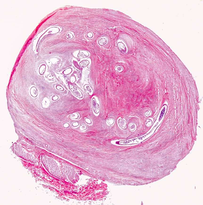
Let’s move to the next filarial worm in this lesson, Onchocerca volvulus, the cause of onchocerciasis (also known as “river blindness”). These adult worms live in the subcutaneous tissues and are transmitted by blackflies of the genus Simulium. Blackflies live and breed in fast-flowing rivers and streams. Consequently, infections most commonly occur in areas close to fast-flowing rivers and streams (unlike LF which is more widespread). Most of the infections occur in Africa (>95%), with some smaller pockets of endemicity in Yemen and in the Venezuela-Brazil border.
")
Figure 8. Age-standardized DALY rates for Onchocerciasis (taken from Global Health Metrics, Open Access)

Figure 9. Blackfly
Life Cycle

Figure 10. Life cycle of Onchocerca volvulus
Pathogenesis & Clinical Presentation
The clinical presentation of onchocerciasis is highly variable and depends on specific transmission dynamics of each endemic focus. Most individuals require repeated exposure to the parasite in order to become infected (less common in travelers). The incubation period is usually in the order of months - typically 12-18 months. While many infections remain asymptomatic, symptomatic cases may involve skin disease, eye disease or both.
Answer
YESSSS. Just like Lymphatic filariasis O. volvulus DOES CARRY Wolbachia - an endosymbiotic intracellular gram negative bacteria. Doxycycline can be used as treatment for onchocerciasis in certain settings.