Other arthropods & ectoparasites
Other arthropods & ectoparasites
Editors: Mónica Pachar, José Antonio Suárez, Laura Naranjo
Section Contents
")
Figure 1. Some ectoparasites of medical importance
Louse (plural: lice)
Lice are 6-legged ectoparasites (live on the outside of the host). They do not jump or fly, and thus transmission among humans is through either: direct contact or use of infested objects (e.g., hair brushes, sharing head caps/hats, sharing towels/clothes/linen). There are three types of lice that affect humans:
- Pediculus humanus capitis (Head lice) → common in school-age children. Infestation characterized by intense scalp pruritus as a result of allergic response to lice antigens. Not vectors for human disease.
- Pediculus humanus humanus (Body louse) → adjusted for the human body. Infestation occurs mainly through clothing and bedding or direct contact; and therefore is more common in individuals under inadequate sanitary conditions (e.g., living in shelters, refugees). Are the main vectors of three pathogens:
- Rickettsia prowazekii (Epidemic typhus)
- Bartonella quintana (Trench fever)
- Borrelia recurrentis (Relapsing fever)
- Phthirus pubis (Pubic or crab louse, or “crabs”) → adjusted for pubic hair and eyebrows. Can be transmitted through sexual contact or via contaminated garments. Infestation characterized by intense pubic pruritus. Are not vectors for human disease.
Treatment: several over the counter topical formulations are available (pyrethrins, permethrin, benzyl alcohol lotion). Oral ivermectin not FDA approved but sometimes used off-label. Most regimens need to be reapplied after 10-14 days as they only kill adults, but not eggs.
| Lice | Preferred treatment | Additional measures | Alternative treatment | Head lice | 1% permethrin cream | Use lice comb | PO ivermectin |
| Body lice | 5% permethrin cream | Environmental decontamination (e.g., changing infested clothes) | PO ivermectin |
| Pubic lice | 1% permethrin cream | Remove visible eggs. Treat sexual partners. | PO ivermectin |

Figure 2. Types of lice

Figure 3. Body lice

Figure 4. Lice bites

Figure 5. Crab louse
Mites
Mites are 8-legged ectoparasites. Are not visible to the naked eye. There are three diseases that involve mites that you should remember:
- Scabies → arguably, the most important and neglected of the three. It is caused by the mite Sarcoptes scabiei var hominis (please see the Scabies module).
- Rickettsialpox → caused by Rickettsia akari. Mites function as vectors between infected rodents and humans. Humans get infected when bitten by mites. Geographical distribution: worldwide, but primarily distributed through urban areas.
- Scrub typhus → caused by Orientia tsutsugamushi. Mites function as vectors between infected rodents and humans. Humans get infected when bitten by mites. Geographical distribution: Asia-Pacific region.

Figure 6. Chiggermite vector of Orientia tsutsugamushi
Myiasis
We'll be spending extra time on myasis because it can be a complex topic. The term “myiasis” refers to the infestation of mammals by fly larvae (maggots). This condition is most often seen in patients with poor hygiene, those from lower socioeconomic background or travelers returning from areas where myiasis is endemic. Depending on the type of myiasis living close to animals can also be a risk factor for human myiasis. There are many species of flies that can cause myiasis; however, we will focus on just a select few. Myiasis can be classified as:

Figure 7. Flies
- Cutaneous myiasis → refers to when maggots infest the skin. There are three types of cutaneous myiasis:
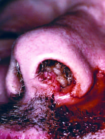
- Furuncular myiasis → occurs when larvae penetrate intact skin forming a furuncle-like nodule with central opening on healthy skin usually with a singular maggot inside. The two most common species are:
- Dermatobia hominis ("human botfly") → is the most common cause of furuncular myiasis in the Americas. Adult flies lay eggs on the abdomen of an intermediary host (ie, mosquito), which when takes a blood meal in mammals, puts them down, liberating larvae that penetrate intact skin.
- (1) Clinical clues → nodules with central opening with surrounding inflammation on previously healthy skin. Lesions are seen in exposed areas. May have pain, pruritus, movement sensation in the lesion (mostly at night), and seropurulent exudate. Sometimes you can see black spiracles (larval hooks).
- (2) Prognosis → larvae cannot complete their full life cycle in humans, so after 5-10 weeks, larvae will emerge and drop to the soil to pupate.
- Cordylobia anthropophaga ("tumbu fly") → most common cause of furuncular myiasis in Sub-Saharan Africa. Adult flies lay eggs on clothing. Risk factors → contact with contaminated clothes (particularly those left to dry in the soil or in bushes - perfect place for oviposition) #WashAndIronYourClothes
- (1) Clinical clues → nodules with central opening with surrounding inflammation on previously healthy skin of unexposed areas (covered in clothes), like the trunk, buttocks, and the thighs.
- (2) Prognosis → If untreated, larvae will exit the lesion after 2-3 weeks to continue its life cycle.

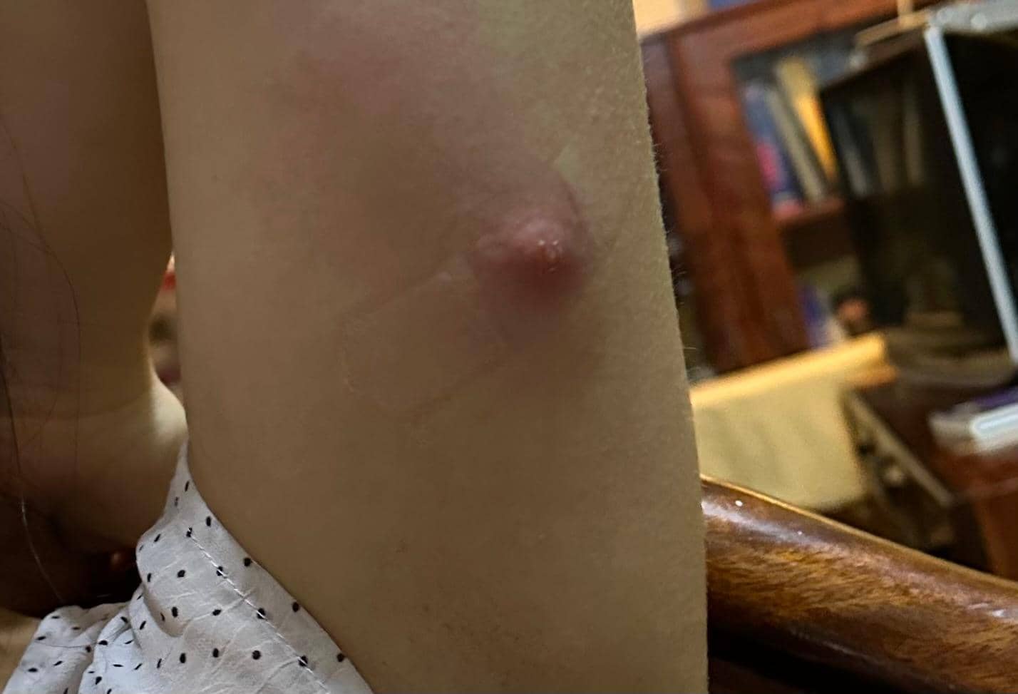
Figure 8. Furuncular myasis in a traveler coming from Uganda (courtesy of Laura Aponte, MD)
Video 1. Furuncular myasis in a traveler coming from Uganda (courtesy of Laura Aponte, MD
- Dermatobia hominis ("human botfly") → is the most common cause of furuncular myiasis in the Americas. Adult flies lay eggs on the abdomen of an intermediary host (ie, mosquito), which when takes a blood meal in mammals, puts them down, liberating larvae that penetrate intact skin.
- Wound myiasis → occurs when an adult flies directly lays eggs in previously open wounds (for example, recent surgery, recent arthropod bites, skin/oral cancer) or in mucous membranes (ie, patients sleeping with mouths open) - opportunistic! In contrast with furuncular myiasis, there are typically multiple maggots in one site. Maggots feed on healthy tissue and thus pre-existing wounds typically enlarge, and may extend into deeper tissue planes. Larvae may be visible in open wounds or mucous membranes. These larvae are called screwworms because of the ability to screw through tissues. The most common species are:
- Cochliomyia hominivorax → more common in the Americas. It affects livestock more frequently than humans.
- Chrysomya spp → more common in the old world (Africa, Indian subcontinent, Southeast and East Asia).
- Migratory myiasis → larvae penetrate skin, form tunnels in the epidermis, and migrate through skin. Sometimes forming serpentine-like burrows, which might be painful and can induce peripheral eosinophilia. Among the differential diagnosis for migratory cutaneous lesions with eosinophilia is: gnathostomiasis, sparganosis, and cutaneous larva migrans
Note: Unlike cutaneous larva migrans, lesions in migratory myiasis can persist for longer periods of time. The most common agents are: Gasterophilus spp ("horse bot fly") and Hypoderma spp ("cattle bot fly").
- Furuncular myiasis → occurs when larvae penetrate intact skin forming a furuncle-like nodule with central opening on healthy skin usually with a singular maggot inside. The two most common species are:
- Cavitary myiasis → occurs when infestation occurs in natural body cavities. Eggs can be deposited in nasal or oral mucosa, conjunctiva, urogenital tract, trachea, or might be accidentally ingested and larvae can penetrate intestinal tract. There is an association with ocular myiasis and flies of the genus Oestrus. Cavitary myiasis can happen with any of the species that cause cutaneous myiasis, as well as with many others.
Figure 9. Myasis
Note: Medical debridement therapy is an important concept you may come across in clinical practice. Although this is not a classification of myiasis, but rather a therapeutic use of fly larvae. Flies of the genus Lucilia are FDA-approved as sterilized maggot therapy in the management of chronic non-healing wounds that have not responded to standard therapy.
- Diagnosis: regardless of the type of myiasis, diagnosis and management have similar principles. Diagnosis is typically clinical as identifying the larvae is the gold standard. Additional imaging (e.g., CT, MRI) may help delineate extent of the lesion if type of myiasis crosses tissue planes.
- Treatment: remove the larvae. Physicians may use oxygen obstructing techniques (e.g., petroleum, mineral oil) to the wound which prompts the larvae to come out to seek for oxygen, so it can be mechanically removed. Oral ivermectin use is off-label and may kill and leave retained larvae inside. Avoid squeezing lesions as it can destroy maggot, and retained antigens may induce a granulomatous response.
Answer
- Migratory myasis
- Gnathostomiasis
- Sparganosis
- Cutaneous larva migrans
Fleas
Fleas are six-legged ectoparasites, meaning they live on the external surface of their hosts. They reach their hosts by jumping. Fleas are medically important because they are involved in the transmission of several human diseases. Clinically, flea bites typically present as papular skin lesions. When it comes to fleas that act as disease vectors, we will concentrate on three medically-relevant species:
- Ctenocephalides felis/canis (“cat/dog flea”) → are the most common fleas in human households. Are involved in the transmission of various diseases:
- Bartonella henselae (Cat-scratch disease)
- Intermediate host of cestodes (ie, Hymenolepis nana, Dipylidium caninum)
- Xenopsylla cheopis (“rat flea”) → is the main vector for:
- Yersinia pestis (Plague)
- Rickettsia typhi (Murine/Endemic typhus)
Figure 9. Myasis
- Tunga penetrans (“sand flea”) → different from other fleas that function as disease vectors, Tunga spp. -by itself - causes the disease tungiasis. It occurs in South America, West Indies & Africa. The flea lives in the soil, and penetrates the skin (usually in the feet while walking barefoot), and causes a characteristic nodular lesion with a central black spot. It might be complicated with tetanus, bacterial infections, or gangrene. Diagnosis is clinical. Treatment involves removal of the flea.
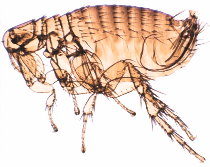

Figure 11. Tungiasis
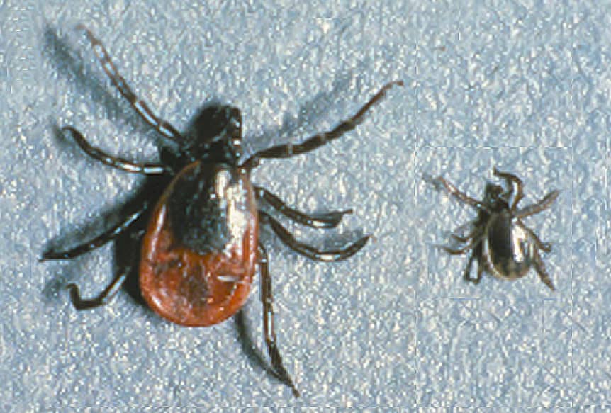
Ticks
They are eight-legged ectoparasites. Learning about ticks can feel overwhelming because they can be classified in multiple ways and are involved in the transmission of a wide range of human diseases - including those caused by viruses, protozoa, and bacteria. One common method of classification divides ticks into soft and hard ticks, based on the characteristics of their exoskeleton.
The ones you need to know are:
- Soft ticks - remember the genus Ornithodoros, which can transmit Borrelia spp. (Tick-borne relapsing fever).
- Hard ticks (more common) - please focus on these four ticks:
- Ixodes spp. → among the diseases that can transmit include:
- Borrelia burgdorferi (Lyme disease)
- Anaplasma phagocytophilum (Anaplasmosis)
- Babesia spp. (Babesiosis)
- Powassan virus (it is a Flavivirus and a cause of encephalitis)
Figure 12. Ixodes spp
- Hyalomma spp. → is the principal vector for:
- Crimean-Congo Hemorrhagic Fever virus (CCHFV)
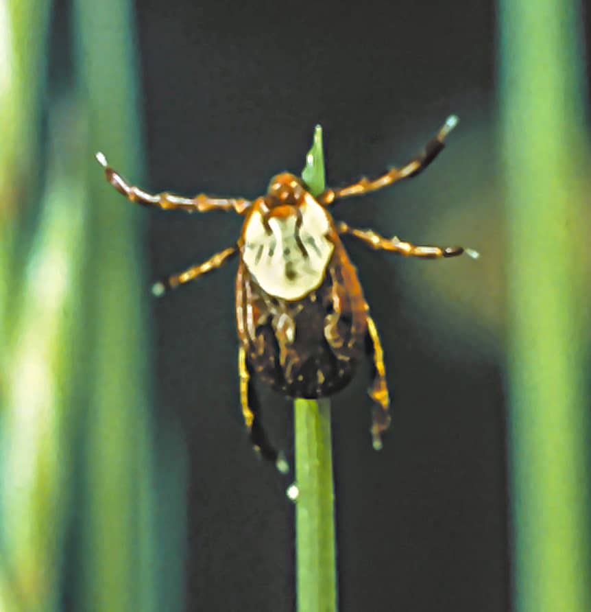
- Dermacentor spp. → among the diseases that can transmit include:
- Rickettsia rickettsii (Rocky Mountain Spotted Fever)
- Francisella tularensis (Tularemia)
- Tick paralysis → caused by a neurotoxin found in a tick's saliva. Produces an ascending paralysis (can be similar to Guillain-Barre syndrome), and can lead to death. Treatment: removal of the tick.
Figure 13. Dermacentor spp
- Amblyomma spp. (Lone Star Tick) → among the diseases that can transmit include:
- Francisella tularensis (Tularemia)
- Ehrlichia ewingii/chaffeensis (Ehrlichiosis)
- Alpha-gal syndrome → is an acquired allergy to meat products (ie, pork, beef, lamb). It occurs after a tick bite which can introduce alpha-gal (a carbohydrate). The body produces an IgE response where subsequent meat consumption can trigger a type I hypersensitivity response.
Bed Bugs
Bed bugs (or Cimex spp) are one of the most frequent ectoparasites that affect humans. They frequently hide in mattresses, box springs, furniture, bedding, walls, floors and personal belongings (e.g., luggage). From here, they crawl and bite susceptible hosts. They induce a papular pruritic rash in a linear pattern (clinical clue!). Aside from infestation and rash, bed bugs do not transmit diseases in humans.

Figure 14. Bed bug
| Arthropods | Genus-Species | Association |
| Lice | Pediculus humanus capitis | Head lice/pediculosis capitis |
| Pediculus humanus humanus |
|
|
| Phthirus pubis | Pubic lice/pediculosis pubis/crabs | |
| Flies (myiasis) | Cochliomyia hominivorax | New World Wound Myiasis |
| Dermatobia spp | New World Furuncular Myiasis | |
| Chrysomya spp | Old World Wound Myiasis | |
| Cordylobia spp (“tumba fly”) | Old World Furuncular Myiasis | |
| Lucilia spp | Maggot Debridement Therapy | |
| Oestrus spp | Cavitary Myiasis, Ocular disease | |
| Flies (non-myiasis) | Simulium spp | Onchocerca volvulus (Oncocercosis) |
| Sandflies (Lutzomyia) |
|
|
| Mites | Sarcoptes scabiei var hominis | Human scabies |
| Chiggers | Orientia tsutsugamushi (Scrub Typhus) | |
| House mites | Rickettsia akari (Rickettsialpox) | |
| Fleas | Ctenocephalides canis (“Dog Flea”) |
|
| Ctenocephalides felis (“Cat Flea”) | ||
| Tunga penetrans | Tungiasis | |
| Xenopsylla spp |
|
|
| Ticks | Ixodes spp |
|
| Amblyomma spp |
|
|
| Dermacentor spp |
|
|
| Hyalomma spp |
|
|
| Ornithodoros spp |
|
Table 1. Summary of medically relevant arthropods
Other Media Resources (Optional)
References

This lesson was built in partnership with Infectotrópico and was last updated August 22 2025