Larval cestodes (echinococcosis)
Larval cestodes (echinococcosis)
Editors (alveolar echinococcosis): David Waldner, Stephen D. Vaughan
Section Contents
- Key Learning Points
- Cystic Echinococcosis (Hydatid Disease)
- Background
- Epidemiology & Transmission
- Life Cycle & Pathogenesis
- Clinical Presentation
- Diagnosis
- Treatment
- Alveolar Echinococcosis (AE)
- Key Differences: CE vs AE
- Summary
- References
Key Learning Points
- Two distinct diseases: Cystic echinococcosis (CE) caused by E. granulosus and alveolar echinococcosis (AE) caused by E. multilocularis have different geographic distributions, growth patterns, and prognoses.
- Silent progression: Both forms can remain asymptomatic for years to decades; CE cysts grow 1-5 cm annually, while AE infiltrates like a malignancy.
- Imaging is cornerstone: Ultrasound WHO-IWGE classification guides management; serology supports but cannot exclude diagnosis.
- Stage-specific approach: Treatment ranges from observation for inactive cysts to surgery for complicated cases; AE requires aggressive therapy.
Cystic Echinococcosis (Hydatid Disease)
Background
Cystic echinococcosis (CE) is the zoonotic infection of humans caused by the larval stage of the cestode Echinococcus granulosus - a parasite that is fundamentally transmitted between dogs and sheep and other livestock. Humans act as dead-end (intermediate) hosts.
Epidemiology & Transmission
E. granulosus is widely distributed, with transmission occurring in pastoral communities and also some peri-urban and urban areas of endemic regions, particularly in South America (Peru, Chile, Argentina, Uruguay), the Mediterranean basin, Eastern Europe, Central Asia, China, and East Africa. In North America, cases can occur in Canada, Alaska, and southwestern U.S. sheep-raising areas; however, most U.S. cases are diagnosed in immigrants.
The adult form of E. granulosus lives in the intestine of domestic dogs and wild canids, and humans become infected through the ingestion of eggs present in canid feces (mainly dog poop). Eggs can survive >1 year in the ideal environment. Children face the highest risk due to poor hygiene and close contact with dogs.
To look at a map showing the endemic area for echinococcosis, can refer to this article: (PDF) Review on Epidemiology and Public Health Significance of Hydatidosis
Life Cycle & Pathogenesis
Summary: Adult tapeworms (2-7mm) develop in the canine small intestine within 32-80 days after ingesting cysts. Gravid proglottids release infectious eggs immediately into the feces. Following human ingestion, oncospheres penetrate intestinal walls, entering portal circulation: 70% lodge in liver, 20% in lungs, 10% reach systemic organs.
Answer
Hydatid cysts develop over the course of months to years, forming complex structures with multiple layers. The innermost layer, the germinal layer, is the biologically active part of the cyst (think like stem cells!), producing hydatid fluid, protoscolices, and brood capsules (vesicles). Surrounding this is the laminated membrane, an acellular structure produced by the parasite that serves as a protective barrier from the host's immune response. Outside of this is the adventitial layer (or pericyst), which is host-derived - composed of fibrous tissue, and sometimes calcifications. This layer walls off the cyst from surrounding tissues.
With time the cyst enlarges, and it may compress nearby organs. Complications include biliary obstruction, portal hypertension, and cyst rupture or leakage, which can trigger anaphylaxis or secondary dissemination. In some cases, fistulas may form, allowing the cyst to drain into the biliary system or lungs.
Over time, the cyst may begin to develop daughter cysts (secondary cysts that grow within the original “mother” cyst) or it may start to degenerate, becoming more solid and calcified. This whole process is dynamic, rather than a staged process.

Figure 1. Life cycle of hydatic cyst.
")
Figure 2. Woman butchering a sheep. The organs containing hydatid cysts are occasionally fed to their dos (completing the life cycle)

Figure 3. Petri dish filled with daughter cysts of E. granulosus
Clinical Presentation
Most infections remain asymptomatic for years; 50% of detected cases are incidental findings. Single-organ involvement occurs in 85-90% with solitary cysts in > 70%. Cysts typically grow 1-5 cm yearly with variable rates.
Diagnosis
Diagnosis starts with a compatible clinical syndrome in the right epidemiological context. Diagnosis of echinococcosis is fundamentally made through imaging and is supported by serology. In the U.S., the CDC can be contacted for diagnostic assistance.

Figure 5. Hooklets of a protoscolex

Figure 6. Protoscolex of E. granulosus

Figure 7. Imaging of a liver infected with multiple daughter cysts

Figure 8. Ultrasound scan of a CE2 (adjacent daughter cysts) cyst in the liver

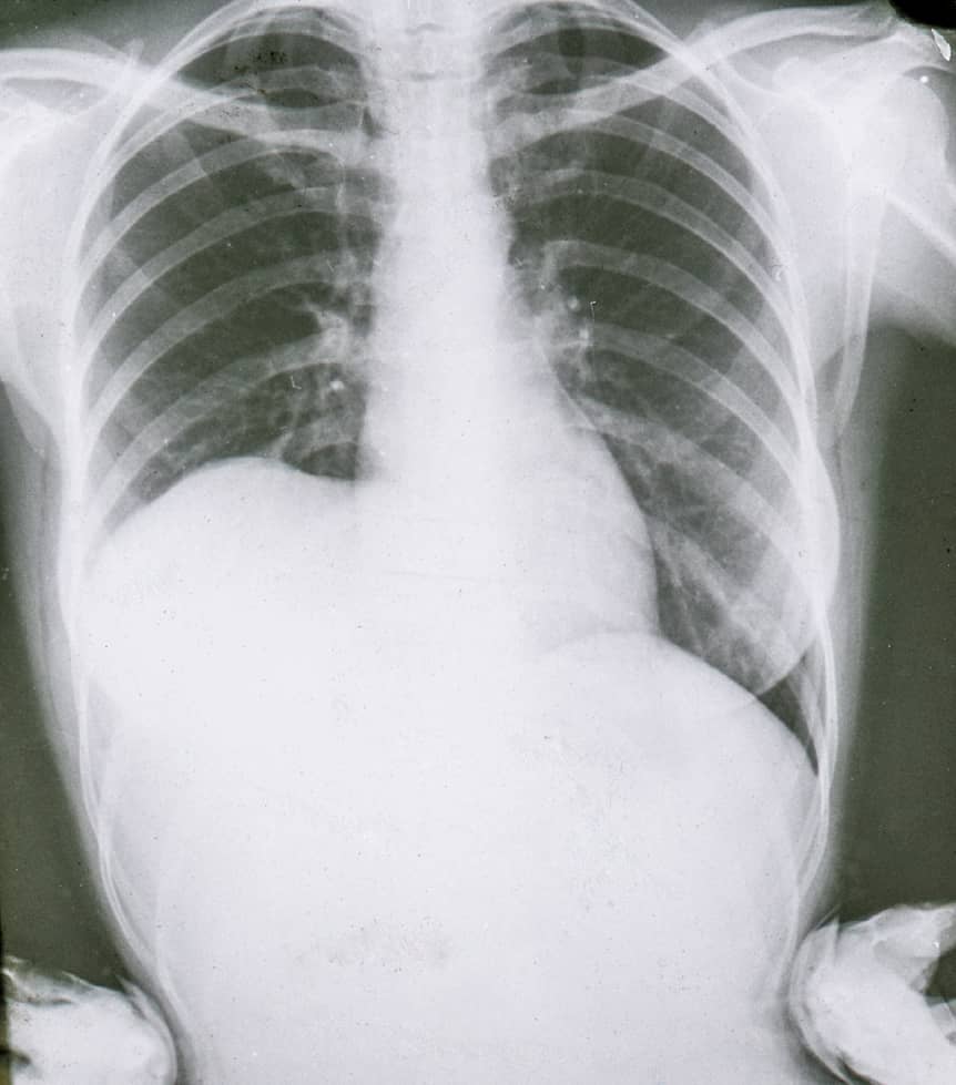
Figure 9. Coronal view of CT scan showing a large echinococcal cyst in the left lung and another echinococcal cyst in the liver (courtesy: Pietro Rinaldi, MD)

Figure 10. CT scan of the chest showing CE3a (note folded endocyst) cyst in the right lung (courtesy: Rafaella Lissandrin, MD)

Figure 11. CT scan of the chest showing small echinococcal cyst in the right pleura (courtesy: Pietro Rinaldi, MD)
Treatment
Treating echinococcus is quite hard. There are guidelines published by the WHO that you can consult here:
However, as a general rule you should remember the following:
- There are four main strategies: 1) “watch and wait” approach, 2) medical therapy (e.g., albendazole), 3) PAIR (percutaneous aspiration with scolicidal injection) or 4) surgical removal (usually with concomitant medical therapy).
- Treatment depends on the presence of complications (uncomplicated vs complicated disease). For patients with complications on clinical presentation (e.g., cyst rupture, allergic complications, bleeding) the treatment of choice is typically surgery.
- For uncomplicated disease, the treatment of choice will depend on the availability of resources along with the size and the location of the cyst (hepatic vs pulmonary CE, <5 cm, 5-10 cm, and >10 cm). In higher resources settings, this will include medical therapy, PAIR or surgery. In many endemic areas the percutaneous treatment options are often limited.
Note: This is not a comprehensive list, but rather a starting point to understand the treatment and management of cystic echinococcosis
- Watch-and-wait: CE4-5 inactive cysts with 6-12 month monitoring
- Medical: Albendazole x 3-6 months; for small CE1 cysts
- PAIR: Percutaneous aspiration with scolicidal injection; consider in CE1/CE3a
- Surgery: For complicated, superficial, or CE2/CE3b cysts or lung cysts.
Alveolar Echinococcosis (AE)
AE, caused by E. multilocularis, occurs across the Northern Hemisphere from Europe to Japan, north-central North America. Maintains the sylvatic cycle between foxes (coyotes in NA) and voles with dogs as aberrant hosts.
Unlike CE's discrete cysts, AE forms infiltrative multilocular masses that invade the liver like a malignancy. After 10-15 years of latency, presents with malaise, weight loss, jaundice—often misdiagnosed as hepatocellular carcinoma. Primary extrahepatic disease is rare (1%), metastases occur in 13%. Untreated mortality >90% at 10-15 years.
Diagnosis shows irregular infiltrative lesions with necrosis on imaging (WHO PNM classification). Patients often undergo multiple biopsies prior to establishing a diagnosis of AE, so a high index of suspicion is required. Pathology shows necrotizing granulomatous inflammation and requires a request for PAS staining (not routinely performed) for identification (or PCR). Emi specific serology supports the diagnosis. Management requires radical resection when feasible plus 2 years of albendazole post-operatively. Inoperable cases require lifelong albendazole. No percutaneous treatment. Modern therapy achieves 88% survival.

Figure 12. Life cycle of E. multilocularis
Key Differences: CE vs AE
| Feature | Cystic Echinococcosis | Alveolar Echinococcosis |
| Distribution | Global pastoral regions | Northern Hemisphere |
| Host Cycle | Dog-sheep | Fox-rodent |
| Growth | Expansile cysts | Infiltrative masses |
| Primary Organ | Liver (67%), lungs (25%) | Liver (>95%) |
| Imaging | Well-defined cysts | Irregular infiltrative |
| Treatment | Stage-specific, including PAIR | Surgery (if possible) + albendazole |
| Untreated Mortality | Variable | >90% at 10-15 years |
Summary
Echinococcosis encompasses two diseases requiring different approaches. CE typically presents as slow-growing cysts amenable to stage-specific treatment. AE behaves as parasitic cancer requiring aggressive therapy. Success depends on early detection through imaging, appropriate staging, and specialized treatment. Prevention through integrated animal control remains most cost-effective for endemic communities.
References
This lesson was last updated October 23 2025