Esquistosomiasis
Esquistosomiasis
Section Contents
Key Points
- Schistosomiasis is a parasitic infection caused by the blood trematode Schistosoma, and it is acquired when larval forms (in freshwater) penetrate the skin.
- Six species are known to cause human disease, but the biggest burden is due to: S. mansoni, S. haematobium & S. japonicum.
- Unlike many other helminthiasis, most of the disease pathology is caused by the eggs and larval stages, and NOT from the adults.
- Diagnosis is supported by microscopy, serology, or imaging.
- Drug of choice for adult worms is praziquantel.
Background
Schistosomiasis (also known as “bilharzia”) is VERY COMPLEX! Please bear with us! This is a parasitic infection caused by the blood trematode of the genus Schistosoma. Schistosomiasis is a neglected tropical disease disproportionately affecting those in rural and impoverished communities in tropical and subtropical areas. Although around 200 million people are estimated to be infected, this number is likely an underestimation since most people have little to no symptoms and the diagnostic methods we have are not that great.
The most important species in humans are: S. mansoni, S. haematobium, and S. japonicum. Other species (S. mekongi, S. intercalatum, and its sister species S. guineensis) play a secondary role on the global burden of schistosomiasis and will only be briefly described in this module.
| Main clinical syndrome | Genus and species | Distribution |
| Urogenital schistosomiasis | S. haematobium | Africa and Middle East |
| Intestinal schistosomiasis | S. mansoni | Africa, South America (Brazil, Venezuela, Suriname) & The Caribbean (Saint Lucia) |
| S. japonicum | South East Asia (China, Philippines, Indonesia) |
|
| S. mekongi | South East Asia (along Mekong River in Laos & Cambodia) |
|
| S. guineensis &
S. intercalatum |
Africa (West & Central) |
Table 1. Summary of clinical phenotypes of schistosomiasis and distribution by species

Transmission & Life cycle
Transmission: infection starts when humans come in contact with stagnant freshwater (never saltwater) containing larval stages (cercariae) and snails (intermediate hosts).
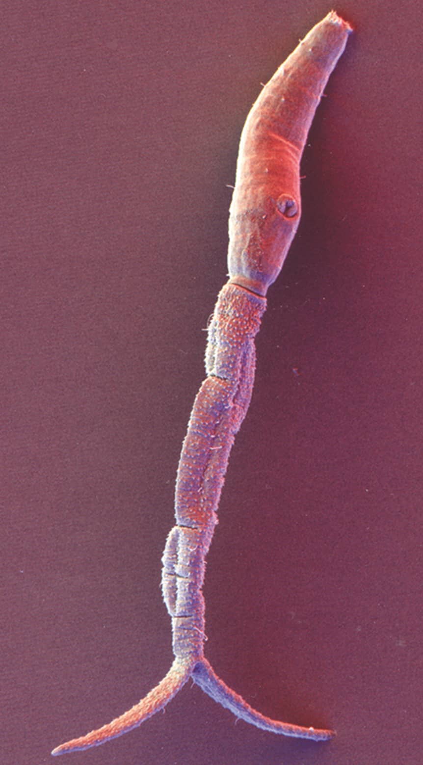
Life Cycle (general): cercariae penetrate the intact skin, and transform into another larval stage called schistosomulae. After ~2 days, schistosomulae migrate through the lungs and mature into adult worms in the portal system of the liver. The disease caused by the transit of schistosomulae through the body is called acute schistosomiasis. Mature adults "crawl" against the blood flow to its definitive habitat in a venous plexus [the type of venous plexus is different for each species- summarized in Table 2]. Once in the definitive habitat, adult females release eggs - which can be swept away to other organs (e.g., liver, intestine, bladder, genital tract) OR can penetrate the intestinal or vesical wall, allowing them to get released with feces (intestinal schistosomiasis) or with urine (urogenital schistosomiasis). After adults start producing eggs, the disease is referred to as chronic schistosomiasis. Eggs passed in feces or urine, hatch in freshwater, and release a larval stage called miracidia, which is capable of infecting the snail [intermediate] host, which is different for each species (please see Table 2). In the snail, miracidia transform back to cercariae, which can infect the human host, and re-start the cycle. Click on the figures below to see the life cycle for each species.

Figure 1. Life cycle of S. mansoni and S. japonicum

Figure 2. Life cycle of S. haematobium
| Schistosoma spp | Intermediate host (snails) | Definitive venous plexus | Reservoirs | Distribution |
|---|---|---|---|---|
| S. haematobium | Bulinus spp. | Vesical plexus and veins draining the ureters | Monkeys | Africa and Middle East |
| S. mansoni | Biomphalaria spp. | Inferior mesenteric veins | Monkeys (Africa), Rodents (America) | Africa, South America (Brazil, Venezuela, Suriname) & The Caribbean (Saint Lucia) |
| S. japonicum | Oncomelania spp. | Superior mesenteric veins | Dogs, Cattle, Rodents | South East Asia (China, Philippines, Indonesia) |
Table 2. Main differences betwen Schistosoma species
Epidemiology
Schistosomiasis is distributed across tropical and subtropical areas; although >85-90% of cases worldwide occur in Africa. For a map of the endemic areas of bilharzia, we recommend accessing this excellent article!
Risk factors for acquiring schistosomiasis:
- Lack of access to clean water & poverty
- Exposures to bodies of freshwater (e.g., washing clothing or dishes, recreational contact,
showers directly connected to lakes, work-related: agriculture, rice farmers) - Encroachment into irrigation canals
- Living in close proximity to water
- Children
Pathophysiology
Unlike many other helminthiasis, the pathology is NOT caused by the adult worms, but rather by the eggs or the larval stages. Let's understand how schistosomes cause disease:
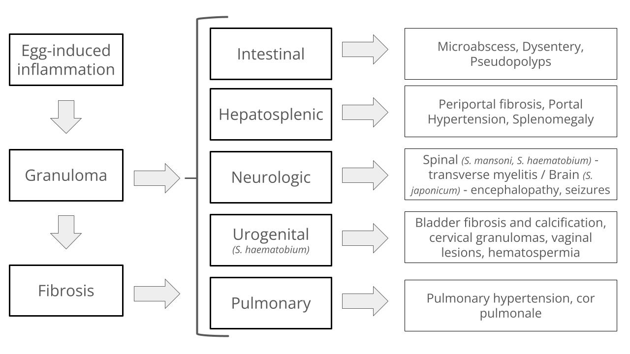
Clinical Presentation
You're almost done with this lesson! Keep up the good work! One of the most challenging features of schistosomiasis is the many different phenotypes it can cause, and symptoms will depend on the stage of the disease:
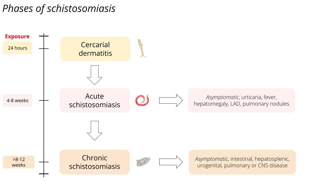
Figure 4. Phases and timeline of schistosomiasis
Diagnosis
As with all diseases, diagnosis starts with a clinical suspicion in the right epidemiological context (What is the presentation? Who is the host?). Definitive diagnosis can be supported through different modalities based on which phase of schistosomiasis you are suspecting:

Figure 5. S. mansoni egg - lateral spine

Figure 6. S. haematobium egg - terminal spine

Figure 7. S. japonicum egg - no spine
")
Figure 8. Schistosoma egg in colon polyp in a patient from Cote D'Ivoire (courtesy of Stephen Lagana, MD)
Treatment & Management
- Swimmer’s itch: symptomatic treatment (e.g.., antihistamines, topical steroids,
emollients) +/- systemic steroids. - Acute schistosomiasis: symptomatic treatment (similar to swimmer’s itch) +
praziquantel.
3. Chronic schistosomiasis:
- All clinical forms (except neuroschistosomiasis) → treat with praziquantel.
- Neuroschistosomiasis → prednisone followed by praziquantel.
| Condition | Treatment |
|---|---|
| Swimmer's itch | Symptomatic treatment +/- steroids |
| Cercarial dermatitis, Acute schistosomiasis | Symptomatic treatment +/- steroids; Praziquantel (may need retreatment) |
| Chronic schistosomiasis | Praziquantel; start with steroids in neuro-schistosomiasis |
Please remember these management pearls:
- Always screen for signs/symptoms of neuroschistosomiasis & neurocysticercosis before treating! Remember praziquantel can induce seizures, myelitis, eye damage in coinfected patients
- In patients with positive microscopy at baseline, perform a test of cure (e.g., stool/urine microscopy) 3-6 months after treatment
- If persistent eggs are seen, re-treat!
- Treatment does NOT prevent re-infection
Prevention
If possible, implement integrated control measures, including:
- Infection avoidance → counsel patients on avoiding recreational swimming in areas at risk, improved sanitation practices (i.e., if possible defecate/urinate in toilets), instruct on safe water practices. If contact with water unavoidable, use personal protection (i.e., boots).
- Health education
- Vector control → snail control and integrated control programs.
- Chemoprevention → praziquantel is given periodically as part of mass drug administration programs in endemic areas with >10% prevalence.
Assessment: Did I Get It? (DIG-IT)
Assessment: Did I Get It? (DIG IT)
DIG ITs are online modules designed to reinforce key learning points for you! Please choose the best answer, then check all of the answer choices for more learning pearls
Other Media Resources (Optional)
References
This lesson was last updated May 12 2025