Babesiosis
Babesiosis
Editor: Luis A. Marcos
Key Points
- Babesia is a zoonotic protozoa that can be acquired through the bite of an Ixodes tick, blood transfusions, vertically-acquired, or through organ transplantation.
- Human babesiosis is primarily seen in North America & Europe and less commonly in Asia.
- Human infection can be either mild/moderate or severe, and it largely depends on the immune status of the host.
- Patients without a spleen or with a non-functioning spleen are at higher risk for severe infection.
- Diagnosis is often a combination of clinical suspicion and microscopy, antibodies, and/or PCR.
- Most patients can be treated with azithromycin + atovaquone.
Background & Epidemiology
Babesiosis is a zoonotic disease caused by protozoa of the genus Babesia, which is primarily transmitted by the Ixodes ticks. The genus Babesia infects the erythrocytes in both humans and a wide array of animals, and ranges from asymptomatic parasitemia to severe multiorgan failure and death.
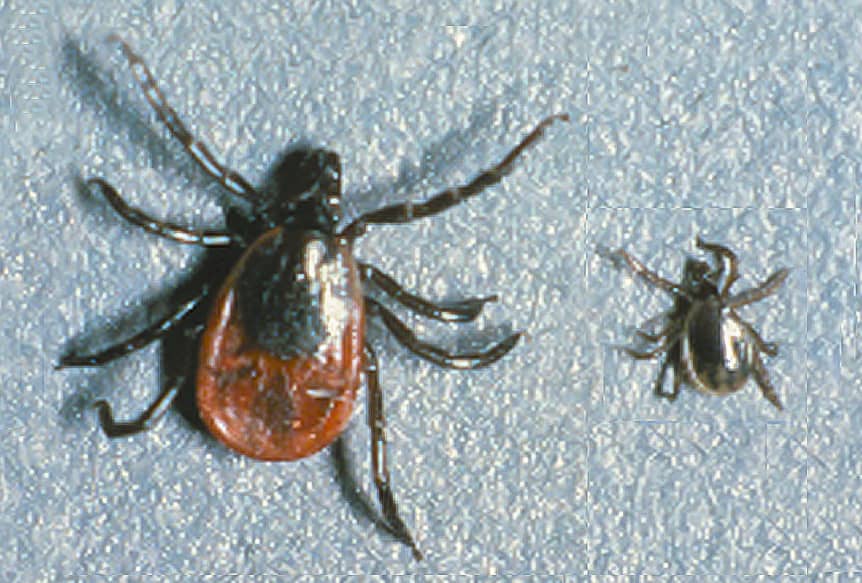
Figure 1. Ixodes spp
Human babesiosis is primarily seen in three regions of the world - summarized in Table 1.
- North America → Babesia is endemic in the Northeastern United States (i.e., Massachusetts, New York, Connecticut), and to a lower degree in the Midwest (i.e., Minnesota, Wisconsin). The dominant species is Babesia microti, which is transmitted by Ixodes scapularis (same vector as Lyme borreliosis and Anaplasma spp). Rodents are the main reservoir (i.e., meadow voles, white-footed mice).
- Europe → Babesia is seen throughout Europe, where is primarily caused by Babesia divergens and Babesia venatorum. These species tend to have a more severe presentation compared to their American counterparts (specially among immunocompromised patients). Cattle is the main resorvoir.
- Asia → much less common than in North America and in Europe. Biggest burden seen in mainland China and to a lesser extent in Japan and Russia. It is caused by the species Babesia microti and Babesia venatorum. Primary vectors are Ixodes persulcatus and Ixodes ovatus.
| Main Distribution | Primary genus/species | Primary vector | Main reservoir | |
| North America | United States (Northeast, MidWest) | Babesia microti | Ixodes scapularis | Small mammals (i.e., rodents) |
| Note → rarely, Babesia duncani, seen in the Western United States | ||||
| Europe | Widely distributed throughout Europe | Babesia divergens, Babesia venatorum | Ixodes ricinus | Cattle |
| Asia | China, Japan, Russia | Babesia microti, Babesia venatorum | Ixodes persulcatus, Ixodes ovatus | Deer (suspected) |
Table 1. Babesia epidemiology
Life Cycle & Pathogenesis
Babesia spp. are closely related to Plasmodium spp. and life cycles share many similarities. Transmission is through an arthropod (i.e., Ixodes tick), but unlike malaria, occurs between animal reservoirs and humans (RARELY between humans). Because Babesia parasites infect erythrocytes, it can also be transmitted through blood transfusions, perinatally or through organ transplantation.
Life cycle: infection starts in humans when tick takes a blood meal and inadvertently introduces sporozoites in the saliva. Sporozoites invade erythrocytes and divide into daughter cells (merozoites) through asexual replication. Merozoites can create ring forms (trophozoites) or tetrad formations (“Maltese cross”), and after replication, they lyse the erythrocyte’s membrane (hemolysis), which releases further merozoites into the bloodstream that are able to parasitize other neighboring erythrocytes. In general, there is no sexual replication (production of gametocytes) in humans, and thus ticks do not continue the life cycle from infected humans.

Figure 2. Life cycle of Babesia spp.
Immunity to Babesia: is complex, and we will not go through it in detail. The most important concept is that the spleen plays a key role in eliminating Babesia-infected erythrocytes. Thus, individuals with immunosuppressive conditions or who have no spleen or a non-functioning spleen may have trouble controlling the infection.
Clinical Presentation
After an incubation period of 1-4 weeks (up to 9 weeks in non-vectorial transmission), babesiosis can present in many different ways. The presentation can vary from uncomplicated to severe, and largely depends on the immune status of the patient. Note that B. divergens & B. venatorum (European forms) are more commonly seen among immunocompromised individuals (therefore, tend to have a more severe course). Click below to see the clinical spectrum of Babesia.
Answer
- FUO or unexplained intermittent fever
- Signs of hemolytic anemia & thrombocytopenia
- Recent exposures → Residence or travel to endemic area (North America, Europe, China), previous blood transfusions, tick bites
- Summer months, less commonly seen in other seasons