Department of Medicine, Division of Infectious Diseases, Columbia University Irving Medical Center, New York, United States
Author: Melissa Parkinson
Carlos Seas MD, FIDSA
Tropical Medicine Institute Alexander von Humboldt. Cayetano Heredia Peruvian University, Lima, Peru (En espanol: Instituto de Medicina Tropical Alexander von Humboldt Universidad Peruana Cayetano Heredia, Lima, Peru).
Leishmaniasis is a parasitic infection caused by the obligate intracellular tissue protozoan Leishmania and is acquired by a bite from a sandfly
Leishmania spp. can be found in many regions of the world and are often categorized into old world (Asia, Africa, and Europe) and new world (the Americas) geographic regions as they have different species that can cause different disease processes
Leishmaniasis can take several forms including cutaneous (CL), mucocutaneous (MCL), and visceral (VL). Visceral leishmaniasis is discussed separately
Definitive diagnosis requires direct identification of the parasite from infected tissue
Treatment can be intra-lesional, oral, or IV, depending on concern for disease progression, the extent of disease, and resources available
Background
Leishmaniasis is a vector-borne zoonosis that encompasses several different disease processes caused by Leishmania spp., and can involve the skin (cutaneous leishmaniasis), the oral, nasal, pharyngeal and/or laryngeal mucosa (mucocutaneous disease), or be a systemic/multiorgan disease (visceral leishmaniasis). Because of its pleomorphism, leishmaniasis can be confusing but don't worry, we’ll try to simplify as much as possible!
Leishmaniasis is considered a neglected tropical disease and primarily occurs in tropical and subtropical regions. The World Health Organization estimates that there are 700,000 to 1 million new cases every year. Cutaneous leishmaniasis, the most common form, is endemic in approximately 90 countries worldwide.
For a more detailed map of the geographic distribution of cutaneous and mucocutaneous disease, please refer to Figure 2 available in the IDSA/ASTMH guidelines.
Figure 1. Clinical syndromes of leishmaniasis
Transmission, Life Cycle & Pathogenesis
Transmission
Leishmaniasis is a vector-borne disease. Leishmania spp. are transmitted through the bite of a female sandfly. In the Western hemisphere (New World) it is transmitted mostly by Lutzomyia spp. and in the Eastern Hemisphere (Old World) it is transmitted by Phlebotomus spp. These bites can go unrecognized as they don’t often hurt or leave mark immediately after the bite. Biting is most prominent from dusk until dawn (night biters!). Sandflies acquire the protozoan after feeding from a mammalian reservoir, and humans are generally incidental hosts - except in a few instances. Animal reservoirs vary based on geographic region.
Life cycle
The life cycle begins when a female sandfly takes a blood meal from an infected mammal and ingests macrophages containing Leishmania amastigotes. The amastigotes develop into promastigotes in the gut of the sandfly. These promastigotes migrate to the proboscis (the straw-like mouth of the sandfly), which are then injected in the saliva into a mammal. The promastigotes are then taken up by macrophages and other mononuclear phagocytic cells, become amastigotes (the tissue stage of the parasite), and multiply in the skin. Infected phagocytic cells are lysed, and inflammatory cells are recruited to the area - leading to the cutaneous lesions over the course of weeks (Figure 2). In the case of species that carry potential for mucosal disease, such as L. braziliensis (more on this later!), the life cycle is similar, with the exception that infected phagocytic cells can leave the primary area of infection and travel to mucous membranes (notably, the nasal, oral and pharyngeal mucosa); where the infection can occur and cause progressive tissue destruction (Figure 3)
Pathogenesis of CL and MCL
An infection with Leishmania can result in a variety of clinical manifestations from asymptomatic infection, self-resolving cutaneous lesions, extensive and destructive mucocutaneous disease, and life-threatening visceral disease (discussed later). Disease and pathogenesis is dependent on the species of Leishmania and host interaction with the parasite.
Figure 2. Life cycle of L. tropica
Figure 3. Life cycle of L. braziliensis
Figure 4. Sandfly
What should you remember regarding the immune response in CL and MCL?
Answer
An important point to remember is that the immune response to Leishmania parasites in both cutaneous and mucosal disease is primarily cell-mediated (Th1). While this response is effective at eliminating the parasite, it also contributes significantly to tissue damage.
In localized cutaneous leishmaniasis (LCL), a Th1 response leads to activation of mononuclear cells, effective parasite clearance, and eventual healing of the lesion. In contrast, mucocutaneous disease is characterized by an exaggerated Th1 response, which results in potent cytotoxicity and severe tissue destruction, and a very low parasite burden (kills all parasites). Cell-mediated immunity is not prominent in all forms of cutaneous leishmaniasis. For instance, diffuse cutaneous leishmaniasis does not follow this Th1-dominant response, and it is primarily mediated by a humoral response (Th2).
Clinical Manifestations
Species
Geographic Distribution
Visceral
L. donovani
Old World (OW)
L. infantum (also known as L. chagasi)
OW and New World (NW)
Mucocutaneous
L. braziliensis
NW
L. panamensis
NW
L. guyanensis
NW
Cutaneous
L. tropica
OW
L. major
OW
L. aethiopica
OW
L. amazonensis
NW
L. colombiensis
NW
L. garnhami
NW
L. guyanensis
NW
L. lainson
NW
L. lindenbergi
NW
L. mexicana
NW
L. naiffi
NW
L. panamensis
NW
L. peruviensis
NW
L. pifanoi
NW
L. shawi
NW
L. venezualensis
NW
L. braziliensis
NW
Diffuse cutaneous
L. aethiopica
OW
L. mexicana complex
NW
L. amazonensis
NW
Disseminated cutaneous
L. braziliensis
NW
Leishmania recidivians
L. tropica
OW
Table 1. Clinical manifestations and associated species in cutaneous and mucosal leishmaniasis
Clinical Presentation and different phenotypes
As mentioned above, there are several different manifestations of disease based on the species and host factors.
Cutaneous leishmaniasis (CL)
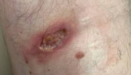
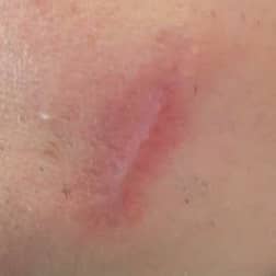
CL presents in several different ways (as noted in Figure 1 and Table 1). We want you to focus on localized cutaneous leishmaniasis (LCL). LCL usually first appears as a red papule 2-8 weeks (but can be up to several months!) after an initial inoculation of promastigotes from the bite of the sandfly and can progress to a nodule and then to a larger, painless, ulcerated lesion (Figure 5) with raised edges, that can crust over and eventually scar. Remember, all lesions leave a well-demarcated atrophic scar! (Figure 6) Classically, cutaneous lesions have raised edges (“volcano edge”), which is where you will find the amastigotes—making sampling this area crucial in diagnosis.
Cutaneous lesions can vary greatly in their presentation and can have smaller satellite lesions around the primary one and be associated with regional lymphadenopathy. Although the ulcerative form is the most common, lesions can be non-ulcerative, including nodular or plaque-like and can be distributed in a lymphangitic or sporotrichoid pattern (quite complex, huh?). To make it even harder, cutaneous lesions can become superinfected with bacteria, which can also alter their appearance.
Note: Like it weren’t complicated enough with LCL, cutaneous leishmaniasis can also take other more rare variants caused by specific species, listed in the table above. We will not describe them in detail, but you should know they exist.
Diffuse cutaneous leishmaniasis (DCL) can occur from several species listed above. It starts out as a single lesion and then can spread over the entire body. Lesions can be papular, nodular or plaque-like, but typically have no visceral or mucosal involvement. DCL is characterized by a Th2 immune response and on biopsy abundant parasites are seen (ineffectively killed by cell-mediated immunity). Treatment is hard, prognosis is poor and relapses are common.
Disseminated cutaneous leishmaniasis (DL) is an aggressive form that presents with several pleomorphic lesions in non-contiguous body regions. Unlike DCL, a Th1 response is more prominent in DL and very few parasites are seen at the lesion. More common in new world disease (e.g., L. braziliensis).
Leishmania recidivans - caused by L. tropica (OW) is a rare chronic/relapsing form with relapsing lesions at the scar site.
Mucocutaneous leishmaniasis (MCL)
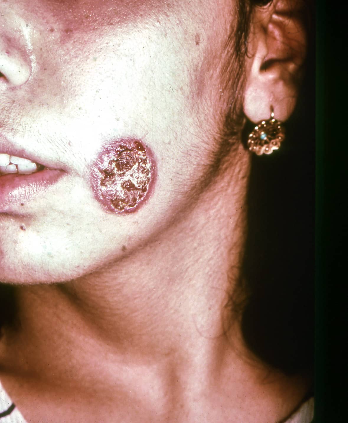
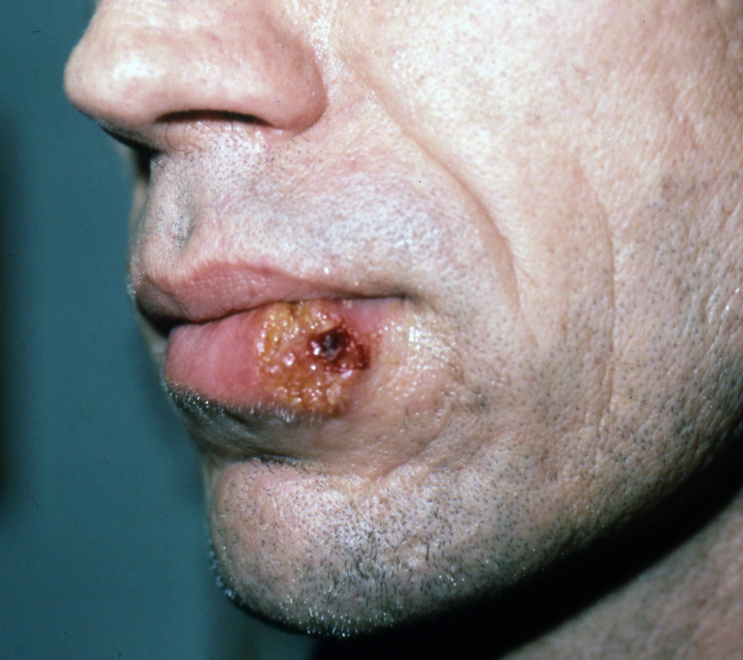
MCL is a complication of the cutaneous form, and it usually occurs months to years (can be decades!) after a primary infection and cutaneous lesions have healed. It can sometimes occur alongside cutaneous lesions. MCL only occurs in the new world and only by select species, most common being L. braziliensis (>95% of cases), L. panamensis, and L. guyanensis. Over 90% of cases occur in Brazil, Bolivia, and Peru. Progression from CL to MCL varies based on species and host factors, with rates about 10-30% of infections progressing to MCL. The parasites spread to mucocutaneous junctions and cause destructive lesions in the nasal, oral, pharyngeal and/or laryngeal regions. Patients can present with nose bleeds, brassy cough, or hoarseness based on the location of the lesions. Severe lesions can be extensive and destructive.
Figure 5. Localized cutaneous leishmaniasis in a patient with L. mexicana. Courtesy of Jorge Cardenas-Alvarez, MD
Figure 6. Localized cutaneous leishmaniasis
Figure 7. Scar following treatment for localized cutaneous leishmaniasis in a patient with L. mexicana. Courtesy of Jorge Cardenas-Alvarez, MD
Figure 8. Mucocutaneous leishmaniasis
Figure 9. Severe mucocutaneous leishmaniasis
Diagnosis
Note: When to suspect cutaneous leishmaniasis? → cutaneous leishmaniasis should be suspected when a person is from or has visited an endemic area and has a skin lesion that fails to heal properly.
When to suspect mucocutaneous leishmaniasis → mucocutaneous leishmaniasis (MCL) should be suspected when a person is from or has visited an endemic area where species causing MCL is known to exist and has destruction of mucosal surfaces concerning for MCL. The patient may recall prior cutaneous lesions that resolved months to years prior.
Diagnostic modalities for cutaneous leishmaniasis
There are several different ways to confirm the diagnosis of leishmaniasis. Identifying the organism directly from the lesion is the best way to confirm diagnosis, though it can be difficult if resources and/or expertise in Leishmania identification is lacking. This is done by taking a sample (scrapings, aspiration, or biopsy) from the edge of a lesion where the parasite is most likely to be present, and sending it for either culture, histology, microscopy (staining with Giemsa!), and/or nucleic acid amplification test (NAAT). Using NAAT for diagnosis is particularly helpful to identify parasite species, which can help you determine if there is risk for progression to MCL, and could change treatment.
In addition, there are two diagnostic tests that can assist, but do not confirm the diagnosis:
Serology: Testing for the presence of antibodies can be done, though it can only determine if the patient has been exposed to Leishmania. This can aid in diagnosis, especially in those from non-endemic areas; although generally is not so helpful.
Leishmaniasis skin test (Montenegro test): much like a TB skin test (TST or PPD), is used in some countries to help in diagnosis. It also only identifies those who have been exposed to Leishmania at some point and cannot distinguish between active or prior infection.
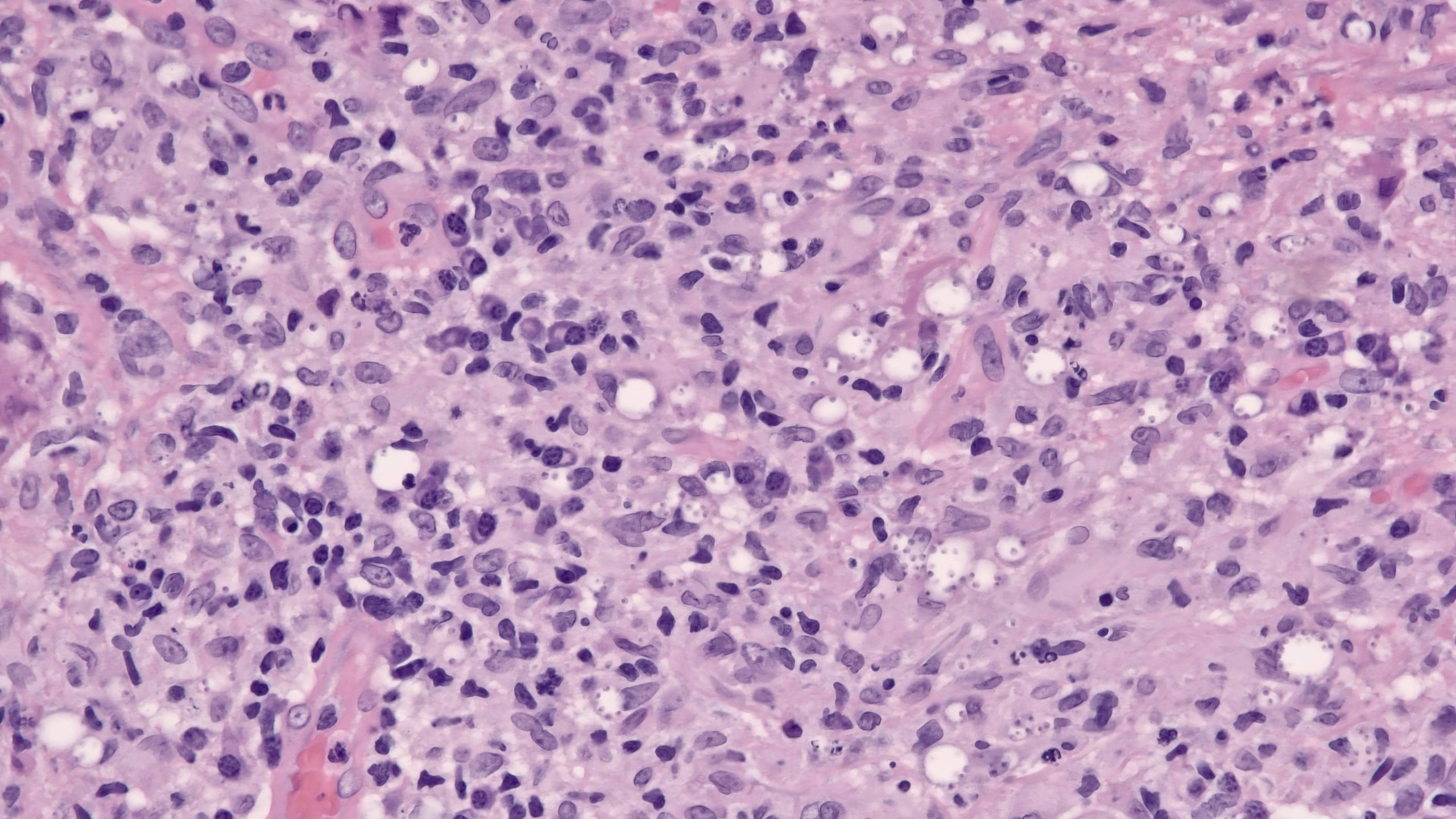
Figure 10. 40X amastigotes with marquee sign (organisms arranged around the periphery of intrahistiocytic vacuoles).
Diagnostic modalities for mucocutaneous leishmaniasis
The same diagnostic tools can be used to confirm MCL, however it is often more difficult to isolate the parasite with direct observation as they are difficult to find in tissue, making NAAT the most reliable tool.
Treatment
Treatment is very hard, and you will need to treat this in consultation with an expert! Treatment of leishmaniasis is dependent on the type of disease, risk of progression, and therapies available. For a more detailed explanation please consult societal guidelines:
Not all lesions require treatment. For example, small, simple/uncomplicated lesions that are not at risk for mucosal disease may be observed as LCL are generally self-healing
If treatment is indicated or requested, there are two therapeutic options – you can either use topical therapy (e.g., cryotherapy, thermal therapy, topical paromomycin) or can use systemic therapy (e.g., IV liposomal amphotericin B, miltefosine)
Choosing one therapeutic agent versus another is done on a case-by-case basis
Treatment of localized cutaneous disease
Generally, local treatment is appropriate for cutaneous leishmaniasis when there isn’t concern for progression to MCL or the lesions aren’t too big, too numerous, or in difficult to treat areas. If you can’t differentiate between species in an area where there are species that can progress to MCL, systemic treatment should be given. This is particularly important in the new world where L. braziliensis is pervasive and overlaps in geographic distribution with Leishmania that only causes cutaneous leishmaniasis. If you are not able to identify the species in this case, it should be treated systemically. It can also be treated with systemic oral or IV therapy if it does not respond to treatment or there are multiple lesions or large lesions that would make local therapy difficult or likely to fail.
Treatment options:
Local therapy: includes physical therapies such as cryotherapy or thermal therapy and topical therapy such as topical paromomycin or intra-lesional pentavalent antimony.
Systemic therapy: include IM or IV pentavalent antimonials, IV liposomal amphotericin, or oral miltefosine.
Treatment of mucocutaneous disease (or at risk for MCL)
Treatment for MCL requires systemic therapy, often for prolonged periods. Systemic therapies include pentavalent antimonials, liposomal amphotericin, or miltefosine.
Prevention
Prevention of sandfly bites will help prevent leishmaniasis. Strategies are similar to other vector-bone diseases. For example, sleeping under bed nets, using insect repellant sprays, wearing insecticide treated clothing, and avoiding activities during peak sandfly activity (early morning or late evening) will help prevent spread
Eradication campaigns of sandflies near highly populated areas, prevention of reservoir infections (for example domestic dogs wearing pyrethroid-impregnated collars) are all additional ways to prevent spread
Vaccines are under investigation, however there are no current vaccines on the market
Visceral leishmaniasis
Key Aspects
Visceral Leishmaniasis (VL), which is caused by specific species of the tissue protozoan, Leishmania, L. donovani and L. infantum (also known as L. chagasi)
It is characterized by fever and hepatosplenomegaly and is fatal in almost all cases without treatment
Background
Visceral leishmaniasis (VL), also known as kala-azar, is a life-threatening disease transmitted by the sandfly. It is caused predominantly by the species L. donovani and L. infantum. Most cases occur in the old world (East Africa and India with the highest prevalence) with the vast majority of new world cases occurring in Brazil. The WHO estimates that 50,000 to 90,000 new cases occur annually. VL is more common in children.
For a more detailed map of the geographic distribution of cutaneous and mucocutaneous disease, please refer to Figure 3 available in the IDSA/ASTMH guidelines.
Transmission, life cycle and pathogenesis
Transmission: VL is transmitted by the sandfly, Phlebotomus spp. in the Old World and Lutzomyia spp. in the New World. In some areas, humans are considered the primary source, while in others there are important reservoirs such as domesticated dogs and rodents.
Life cycle: The life cycle is quite similar to the one described for CL/MCL; except that infected cells do not lead to pathology in the skin or mucosal orifices, but rather spreads to the liver, spleen, and bone marrow.
Figure 11. Life cycle of L. donovani
Clinical Presentation
VL occurs 3-6 months (up to years!) after initial infection with Leishmania. Some infections can be asymptomatic but when symptomatic can manifest in a variety of ways. Patients are usually febrile and characteristically have a double-daily fever. They can develop generalized lymphadenopathy, hepatosplenomegaly (it’s one of the causes of massive splenomegaly!), anemia, low platelets, darkened skin, wasting and are at risk of superimposed infection - secondary to leukopenia. Immunosuppressive conditions can lead to clinical manifestations of latent infections.
Post-kala-azar dermal leishmaniasis (PKDL) → is a rash that some patients develop months to years after treatment of VL. The rash starts out as hypopigmented or erythematous macules and develops into nodules, which contain amastigotes and can spread infection when the individual is bitten by the sandfly. Distribution often in face and trunk
Congenital VL → VL can be acquired congenitally as infected phagocytes can pass through the placenta. The clinical manifestations of congenital VL are similar to a primary infection
Diagnosis
Diagnosis starts with a compatible clinical syndrome (e.g., fever, pancytopenia, hepatosplenomegaly). Diagnosis can be made by either microscopy (with Giemsa staining!), culture or NAAT from either spleen and/or bone marrow aspirates. Other potential specimen sources include: liver biopsy or buffy coat. This means that when suspecting VL, one can send PCR from tissue and from blood! - isn’t that amazing?
Note: One must be very careful as splenic aspiration is a dangerous procedure with risk of severe bleeding and should be avoided if possible.
In addition, K39 antigen immunochromatographic test (point-of-care) is available, but efficacy varies according to the geographical region. Like with other forms of leishmaniasis, more non-specific tests such as serology or skin tests can be done but do not provide a definitive diagnosis.
Treatment
Systemic treatment with amphotericin, pentavalent antimonials, or miltefosine often in combination, are treatment options. Treatment of choice is based on geographic location and availability of medication. Once again, you will need to treat this in consultation with an expert!
Prevention
As with CL and MCL, prevention consists of vector control and protection against sandfly exposure during peak hours. Reservoir control programs have been met with limited success.
Assessment: Did I Get It? (DIG IT)
DIG ITs are online modules designed to reinforce key learning points for you! Please choose the best answer, thencheck all of the answer choices for more learning pearls
Montaner-Angoiti, E., & Llobat, L. (2023). Is leishmaniasis the new emerging zoonosis in the world? Veterinary Research Communications, 47(4), 1777–1799. https://doi.org/10.1007/s11259-023-10171-5
de Vries, H. J., & Schallig, H. D. (2022). Cutaneous leishmaniasis: A 2022 updated narrative review into diagnosis and Management Developments. American Journal of Clinical Dermatology, 23(6), 823–840. https://doi.org/10.1007/s40257-022-00726-8
del Giudice, P., Marty, P., Lacour, J. P., Perrin, C., Pratlong, F., Haas, H., Dellamonica, P., & Le Fichoux, Y. (1998). Cutaneous leishmaniasis due to Leishmania infantum. Archives of Dermatology, 134(2), 193. https://doi.org/10.1001/archderm.134.2.193
Yizengaw, E., Gashaw, B., Yimer, M., Takele, Y., Nibret, E., Yismaw, G., Cruz Cervera, E., Ejigu, K., Tamiru, D., Munshea, A., Müller, I., Weller, R., Cotton, J. A., Chapman, L. A., & Kropf, P. (2024). Demographic characteristics and clinical features of patients presenting with different forms of cutaneous leishmaniasis, in lay gayint, northern Ethiopia. PLOS Neglected Tropical Diseases, 18(8). https://doi.org/10.1371/journal.pntd.0012409
Scott, P., & Novais, F. O. (2016). Cutaneous leishmaniasis: Immune responses in protection and pathogenesis. Nature Reviews Immunology, 16(9), 581–592. https://doi.org/10.1038/nri.2016.72
Olivo Freites, C., Gundacker, N. D., Pascale, J. M., Saldaña, A., Diaz-Suarez, R., Jimenez, G., Sosa, N., García, E., Jimenez, A., & Suarez, J. A. (2018). First case of diffuse leishmaniasis associated with Leishmania panamensis. Open Forum Infectious Diseases, 5(11). https://doi.org/10.1093/ofid/ofy281
Machado, P. R. L., Lago, A., Cardoso, T. M., Magalhaes, A., Carvalho, L. P., Lago, T., Carvalho, A. M., Costa, R., & Carvalho, E. M. (2024). Disseminated leishmaniasis, a severe form of leishmania braziliensis infection. Emerging Infectious Diseases, 30(3), 510–518. https://doi.org/10.3201/eid3003.230786
Volpedo, G., Pacheco-Fernandez, T., Holcomb, E. A., Cipriano, N., Cox, B., & Satoskar, A. R. (2021). Mechanisms of immunopathogenesis in cutaneous leishmaniasis and post kala-azar dermal leishmaniasis (PKDL). Frontiers in Cellular and Infection Microbiology, 11. https://doi.org/10.3389/fcimb.2021.685296